
Magnetic Resonance Imaging (MRI) is one of the most common medical imaging techniques, performed approximately 100-150 million times annually globally. Chances are that you know someone who has had an MRI scan. But what exactly is an MRI? Why do we use it? And what are the risks and benefits of using such an advanced piece of medical technology?
What is Magnetic Resonance Imaging?
Modern MRI can be credited to Dr. Raymond Damadian. In 1969 theorized that magnetic resonance would be an effective way to detect cancer cells. He would later patent, design, and finally build the first fully functional MRI machine in 1977.

Dr. Damadian’s invention of modern MRIs revolutionizes medical imaging by using magnetic fields and computer-generated radio waves to create detailed images of organs and tissues.
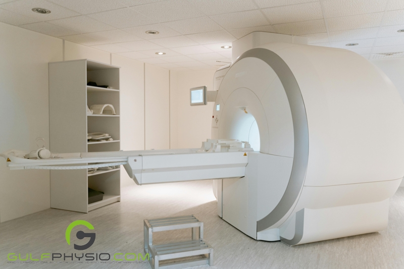
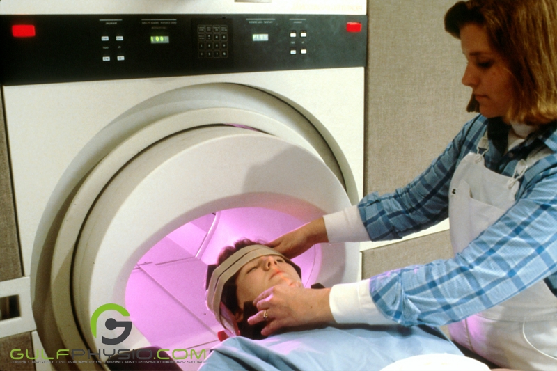
Physically, MRI machines look like giant tubes. The patient is inserted into this giant tube, which is actually a magnet. Once inside the machine, the scan begins and lasts 20 to 60 minutes to complete.
Why do we need Magnetic Resonance Imaging?
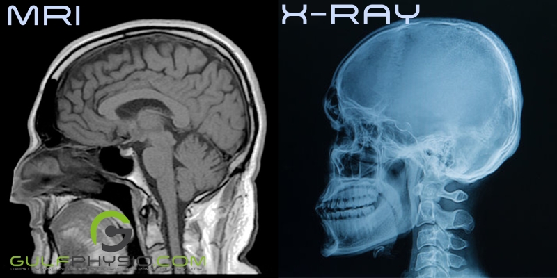
Some of you reading this may be thinking “Why do I need an MRI? Why not just get an X-ray?”. While X-rays are generally cheaper, getting an MRI is more beneficial:

- A safer option: Magnetic resonance imaging (MRI) is a safe medical technique that uses radio waves and magnetic fields to produce images of the body, suitable for all ages, including children and expectant mothers, without the need for contrast agents.

- Noninvasive: MRI is a non-invasive medical imaging method that produces detailed images from multiple angles using magnetic energy pulses. Doctors can get better images without using harsher techniques like biopsies because it does not require needles or incisions.
- More detailed: MRI scans enable physicians to accurately assess the brain, spinal cord, and circulatory system, providing clear images of soft tissue components essential in neurology. They are crucial for identifying brain disorders and providing extensive information about abnormalities.
When should you get an MRI?
The big question is when to ask your doctor for an MRI or which conditions warrant a scan. There are quite a few situations in which magnetic resonance imaging is needed.

- MRIs for breasts – MRI, when combined with breast ultrasound and mammography, can effectively detect breast cancer in women with implants or dense breast tissue, identifying minor lesions missed by mammography. It’s particularly useful for women under 40 and high-risk women.

- MRI for bones – To check for injuries, structural abnormalities, and problems such as tumors, inflammatory diseases, congenital anomalies, osteonecrosis, bone marrow disease, and spinal cord disc degeneration, orthopedic doctors often employ MRI scans. It can also track joint degradation from arthritis and evaluate the outcomes of remedial orthopedic operations.
- MRI for internal organs – Since MRI can differentiate between abnormal and normal soft tissues, it is a more accurate technique than CT scanning for investigating organs. It has been applied to several operations, including: assessing artery blood flow, identifying vascular abnormalities, and identifying brain aneurysms. To pinpoint the exact location of a function, like speech or memory, in the brain, functional magnetic resonance imaging (fMRI) is utilized. A particular task, such as reciting the Pledge of Allegiance (for Americans), is performed during fMRI, and the location of the brain function may differ from person to person. Physicians can use this information to develop therapies, including surgery, for brain disorders.
How To Prepare for an MRI?
It is understandable to feel nervous before an MRI scan, but aside from a few reminders, there is very little preparation required for getting an MRI scan.

- Let them know if you have Claustrophobia – People with claustrophobia may experience anxiety during an MRI since they must lie in an enclosed tube-shaped machine for up to an hour. Talk to your doctor about your claustrophobic history and worries about managing the situation. To keep you calm throughout the treatment, doctors could prescribe medication. To guarantee a seamless encounter, it is crucial to speak with your physician before the surgery.

- Don’t take jewelry with you – Because MRIs are magnets, it is necessary to avoid wearing metal objects, especially jewelry, prior to an MRI. Any metal objects will be asked to be removed by MRI personnel. It could be advisable to leave costly, hard-to-replace jewelry at home. Even though there is little chance that anything will go wrong during the scan, it is always better to be careful than to lose any important accessories.
What to expect during an MRI?
Your comfort during an MRI is crucial. You will receive blankets, earplugs, and a button that will serve as a communication device from your technologist. MRI scans can be done as part of a hospital stay or as an outpatient treatment, and while specific protocols may differ between facilities, the process generally follows the same steps.
The Process
- For certain procedures, such as in the detection of tumors, an IV line will be inserted in the patient’s hand or arm to inject the contrast dye. The contrast will be given to you to swallow if it is to be taken orally.
- The area of your body being scanned, such as your head, knee, or abdomen, may have a coil wrapped around it. This coil, which is typically housed in hard white plastic, is a unique radio receiver that enhances the image. Inform the MRI technologist if the coil starts to cause you any discomfort during the scan.
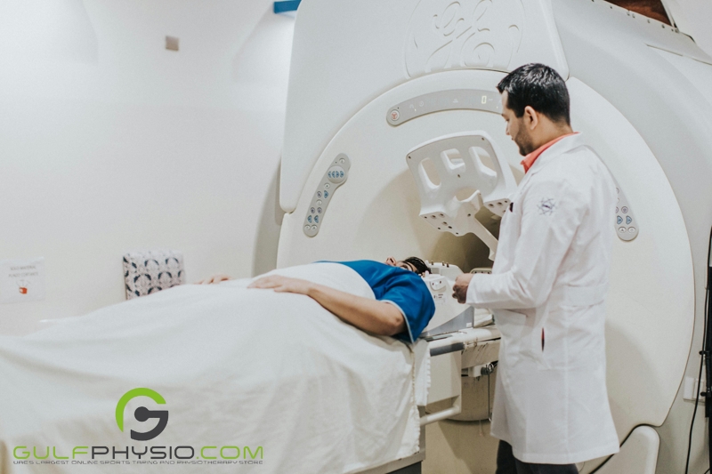
- The patient will be asked to lie on a scan table that slips into the scanning machine’s huge circular hole. Straps and pillows might be utilized to keep the patient from moving while the procedure is being performed.
- You will occasionally hear banging sounds. The shifting magnetic fields are the source of these sounds. The tunnel’s air is moved by fans, so you’ll feel it moving.
- Sometimes, depending on the area being examined, you could be told to hold your breath or to stop breathing for a short while. Then, you will be informed when you are able to breathe. Holding your breath should only be necessary for a few seconds at most.
- The table will glide out of the scanner after the scan is finished, and you will be assisted in getting off the table. The IV line will be withdrawn if one was placed in order to administer contrast.
After the MRI

After an MRI scan, move carefully to prevent lightheadedness or dizziness. Avoid driving and take it easy until the effects of any sedatives have worn off. If contrast dye was used, notify your doctor right away if you experience any negative symptoms or dye reactions. Notify your doctor if you experience any discomfort, redness, or swelling at the IV site, as these symptoms may be signs of an infection. Except for the aforementioned situations, you can resume your regular activities and diet following an MRI scan unless otherwise instructed. Depending on your circumstances, your doctor might give you different or extra directions.
Our Thoughts
Magnetic Resonance Imaging (MRI) is a useful medical tool that can help detect various diseases and disorders without having any exposure to dangerous elements such as radiation. This procedure can provide detailed, three-dimensional images of the organs, providing medical professionals with more information to better provide diagnoses and treatments to patients. Despite this, getting an MRI scan is a serious procedure and may not be a viable option for certain individuals. Consulting your doctor is the best course of action if you are interested in getting an MRI scan
Disclaimer
GulfPhysio.com and all of its content are for informational purposes only. All information is believed to be accurate at the time of posting and should NOT be taken as professional medical advice. Please seek a medical professional in the event of pain or injury.
Want to learn more about health, pain management, and disease? Then read our articles “Tackling the Facts: Transverse Myelitis” and “The Hard-Hitting Reality of Women’s Rugby: Their Battle with ACL Tears”.