Description
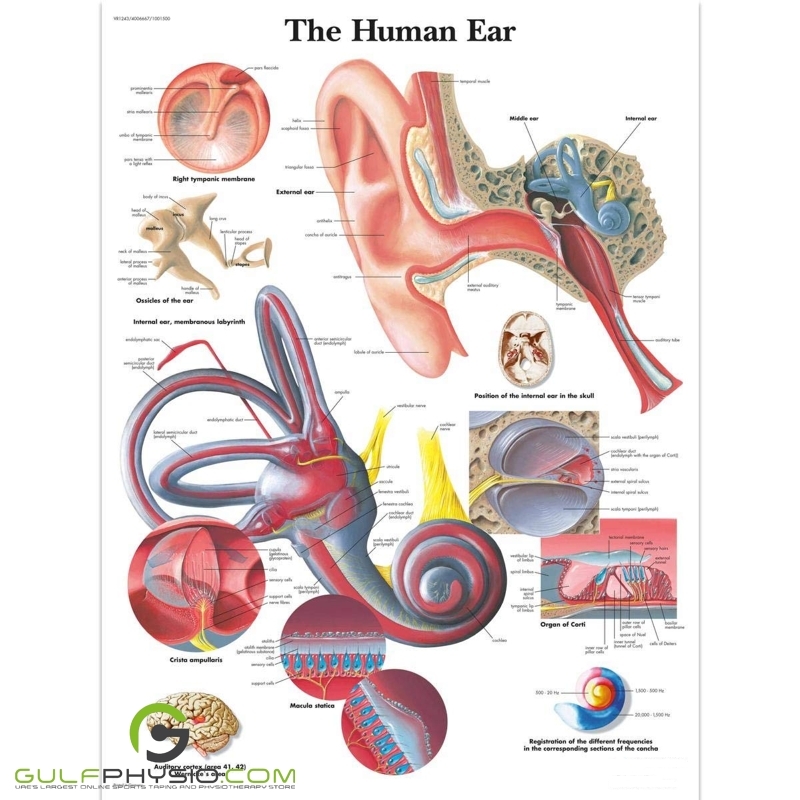
A detailed chart of the anatomy of a human ear.
This anatomical chart details the anatomy of the inner, outer, and middle ear in detail and fully colored. It highlights key structures like ciliated cells, which are located in the cochlea of the inner ear. These specialized cells, with tiny hair-like projections, play a critical role in hearing by converting sound vibrations into electrical signals sent to the brain. This chart is thickly laminated, printed on premium glossy UV-resistant paper, and comes with 2-sided lamination (125-micron, 5.0-mil) and metal eyelets to make the chart easy to display. The 125-micron lamination ensures the chart does not curl up at the edges and the UV treatment ensures the chart does not get a faded yellow color over time.


There are no reviews yet.